Our cardiologists provide a comprehensive cardiac services including consultation, assessment and examination.
Resting Electrocardiography
The 12 lead electrocardiogram measures the heart's electrical activity and records it as waveforms. It is one of the most valuable and commonly used diagnostic examination. It provides basic structural and rhythmic information of the heart.
24 hour Holter Electrocardiogram
(Ambulatory Electrocardiogram)
It allows continuous recording of electrocardiogram as the patient follows his normal routine. It can provide considerably more diagnostic information than a standard resting electrocardiogram and it can detect intermittent and asymptomatic abnormal heart rhythm.
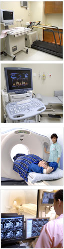
Treadmill Exercise Stress Test
During the test, the patient will walk and run on a treadmill with his electrocardiogram continuously recorded. It is performed to evaluate arterial blood flow to heart muscle during physical exercise, compared to blood flow while at rest. As an exercise test, it also reflects overall physical capacity.
Inadequate blood supply to the heart on exercise due to blockages of coronary vessels will be reflected by changes on the electrocardiogram.
24 hour Blood Pressure Monitoring
(Ambulatory Blood Pressure Monitoring)
It allows continuous blood pressure recording as the patient follows his normal routine. It is especially useful to detect borderline hypertension.
Cardiac Event Recorder
It allows recording of electrical activity of the heart on an ��on-demand' basis when patient feels any discomfort or palpitation over an extended period of time such as a few weeks. It is particularly useful for detecting infrequent and short lasting abnormal heart rhythms.
Tilt Table Test
This is a test to evaluate patients with fainting or syncope attacks. Patients with recurrent attacks of dizziness, with or without the loss of consciousness, which are suspected to be associated with a drop in blood pressure or heart rate, are good candidates for the test. During the test, the patient would be strapped to a tilt table, which will be tilted to different angles to evaluate the response of the blood pressure and heart rate in different table inclination.
Echocardiography
By the use of ultrasound technique, it can study the structures and function of the heart and display them in a two or three dimensional mode. It allows assessment of blood flow velocity and direction and it also evaluates the function of the heart valves for any blockage or leakage.
Transoesophgeal Echocardiogram
With this technique, the echocardiogram is performed with the ultrasound probe inside the esophagus, so that the ultrasound probe can be in close contact with the back side of the heart to obtain a clear view of its structures. This study is particularly useful to assess blood clots and tumors, valvular heart disease and congenital heart disease.
Cardiac Magnetic Resonance Imaging
Cardiac MRI uses radio waves and magnets to create images of the heart. It does not carry any ionizing radiation. It is a very useful and accurate test to evaluate coronary heart disease, heart function, blood perfusion and viability of heart muscles.
Cardiac Computed Tomography Scan
Cardiac CT uses advanced CT technology and computer reconstruction technique to visualize coronary artery anatomy, coronary calcification and plaque formation and other structures of the heart. Compared with conventional catheter-based coronary angiogram, it is less invasive and is more convenient. It can acquire the necessary images within a 5-second breath-hold.
|
 |
 |