Coronary Angiogram and Coronary Angioplasty and Stenting

Coronary angiography is usually performed under local anesthesia. A small catheter (a thin hollow tube with a diameter of 2mm or less) is inserted through the skin into an artery in either the groin or the wrist. Under fluoroscopy (a special x-ray viewing instrument) guidance, the catheter is then advanced to the origins of the coronary arteries. A small amount of radio-opaque contrast is injected into the coronary arteries. The images that are produced are called the angiogram.


Angiographic images accurately reveal the extent and severity of coronary artery blockages. They can guide the doctor to select the appropriate treatment, which may include medications, balloon angioplasty and stenting or coronary artery bypass surgery. The coronary angiogram is currently the gold-standard for the precise quantification of the extent and severity of coronary blockages.


If needed, coronary angioplasty can be performed in the same session. Through a guiding catheter, a guide wire would first be passed through the narrowed segment. Then a balloon catheter would be passed to the site of blockage and be inflated. The atherosclerotic plaque would be compressed against the arterial wall with an increase in the vessel lumen. Finally, a metallic stent would usually be permanently implanted to maintain the result. During the angioplasty process, some adjunctive devices may be needed. Among them, intravascular ultrasound study can provide detailed images of the vessel condition. The latest Virtual HistologyTM system of our centre, which is the first installation in Asia and tenth in the world, can provide detailed analysis of the plaque, thereby assisting the physician to choose the best therapy for the patient and improving the outcome and safety of the procedures.


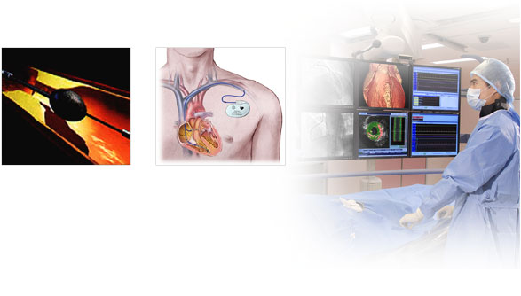
Implantation of Pacemaker / Cardioverter Defibrillator

Pacemaker is a small battery operated computer that can be implanted inside the body to correct slow heart rate problems. The implantation procedure is usually done under local anesthesia and the device is generally implanted under the skin below the collarbone and is connected to the heart via one or more pacing leads.

An implantable cardioverter defibrillator is a device that can generate electrical shocks to correct fatal arrhythmias. It can also act as a pacemaker if slow heart rate is detected. In general, the device is implanted in a similar way as a pacemaker.

Electrophysiological Study / Radiofrequency Ablation

An electrophysiology study is a special catheterization test in which electrode catheters are put inside the heart, usually via the femoral veins in the groins, to study the cardiac electrical system. The procedure is carried out under local anesthesia to check out cardiac arrhythmias.

Radiofrequency ablation is a procedure that utilizes a special electrode catheter to deliver radiofrequency energy to cauterize culprit areas within the heart that are responsible for the arrhythmias. It is carried out in the same procedure, immediately after the baseline electrophysiology study.
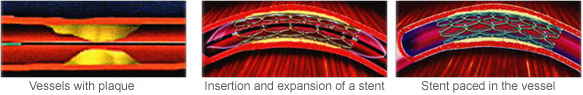